


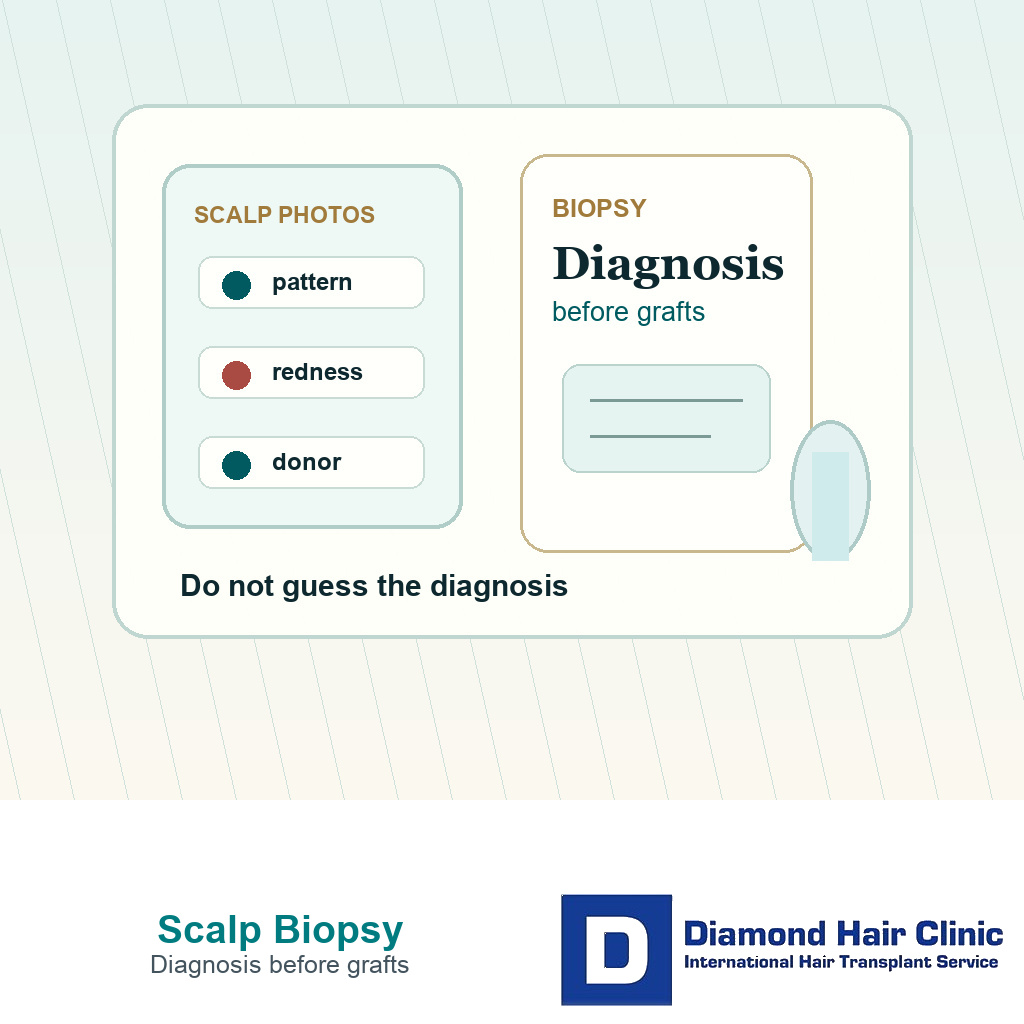
Scalp Biopsy Helps When the Diagnosis Is Unclear
You do not need a scalp biopsy before every hair transplant. I consider it when the diagnosis is still not clear after history, photos, examination, trichoscopy, and basic medical review. A clear androgenetic pattern with a strong donor area and settled scalp often does not need biopsy before a surgical plan can be discussed. An inflamed scalp, diffuse loss, patchy loss, weak donor behavior, symptoms that do not match the photos, or unexplained failed growth changes the level of caution.
I do not use biopsy to make surgery happen faster. I use it, when needed, to avoid the wrong operation. A hair transplant moves hair from one area to another. It does not cure an inflammatory scalp disease, stop an autoimmune pattern, clear infection, or make a weak donor area strong. The diagnosis comes before the graft number.
For that reason, this question belongs inside a proper candidacy discussion, not at the end of a sales conversation. Being a good candidate for a hair transplant depends on a diagnosis clear enough to protect the donor area. A scalp mole or raised spot before a hair transplant belongs in the same category when the skin finding is unclear.
The diagnostic question a scalp biopsy answers
A scalp biopsy is a small skin sample examined under a microscope. In hair loss, it is used when the clinical picture cannot be explained confidently from history, scalp examination, trichoscopy, photographs, blood work, and medication history alone. It can help separate scarring alopecia, inflammatory disease, alopecia areata, telogen shedding, androgenetic miniaturization, or mixed patterns when the surface appearance is not enough.
When biopsy is useful, it answers a tissue question. Is there scarring? Is there active inflammation around follicles? Have follicular openings been lost? Is the pattern mainly miniaturization, infection, shedding, or something mixed? In some uncertain patches, tinea capitis before FUE also needs diagnostic confirmation before cosmetic planning starts. Biopsy does not tell the surgeon how many grafts to implant, and it does not guarantee growth.
The report is evidence, not permission. It still has to be interpreted with the visible pattern, donor area, symptoms, treatment history, and expectations. A reassuring report from the wrong area may not answer the real question. A worrying report may explain why surgery should wait, shrink, or not happen.
Planning without a biopsy in clear cases
If the pattern is classic, the donor area is strong, the scalp is settled, progression is understandable, and there are no signs of scarring or inflammation, biopsy is not routine hair transplant planning. The more useful work is donor measurement, hairline design, graft distribution, medication review, and realistic expectation setting.
Biopsy can also be unnecessary when a dermatologist has already made the diagnosis clearly and the case is stable. Repeating tissue tests without a reason can add a small scar, more anxiety, and no better decision. The useful question is whether anything clinically meaningful is still unknown before donor grafts are used.
I am careful not to use biopsy as a performance of seriousness. A clinic can mention biopsy and still make a poor plan. Another clinic may not need biopsy because the diagnosis is already clear. A test helps only when it answers a real uncertainty in this person.
Reasons I slow down before planning
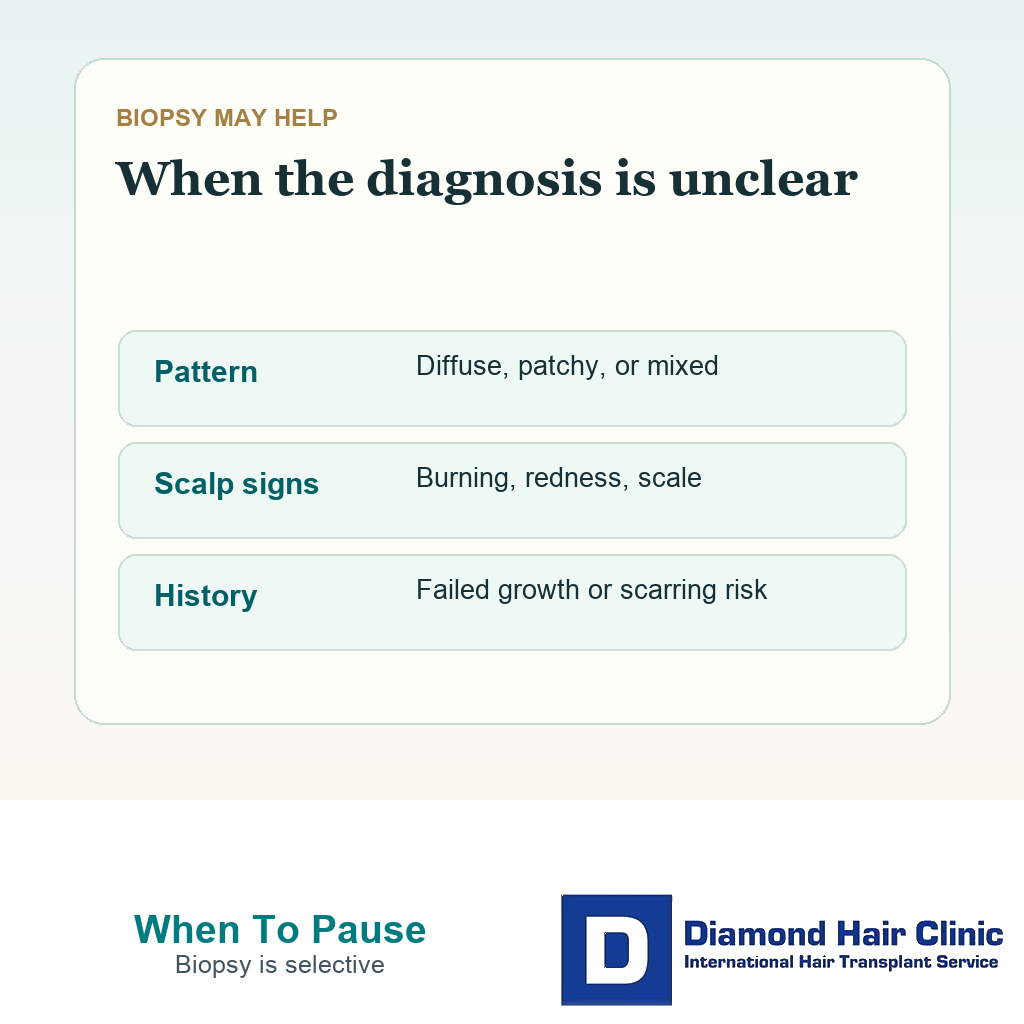
I slow down when the pattern does not behave like ordinary male or female pattern hair loss. Burning, repeated itching, tenderness, scaling, redness, pustules, sudden shedding, patchy loss, shiny scarred skin, loss of follicular openings, eyebrow or beard involvement, or a donor area that looks affected can all change the decision. A radiated scalp can raise a similar tissue question, which is why hair transplant after scalp radiation starts with skin quality rather than graft numbers. Persistent nape bumps are one example, because acne keloidalis nuchae changes donor safety before FUE.
The practical distinction is not one itchy day versus surgery forever. It is whether the symptoms, skin findings, and hair loss pattern repeatedly point away from routine androgenetic alopecia. I also slow down when the story and the photographs do not match. A few images can begin the discussion, but planning a hair transplant from photos should not finish the diagnosis.
Some people arrive after several opinions, multiple medication trials, or a failed transplant with no clear explanation. In that setting, biopsy may be one part of a broader diagnostic reset. It is not a shortcut to surgery. It is a way to decide whether surgery should wait, change, or sometimes not be done.
Biopsy is considered when the uncertainty is diagnostic, not when someone only wants a faster graft number.
Diffuse thinning can mislead planning
Diffuse thinning is one of the places where people move too fast. The scalp may still contain many native hairs, but those hairs may be weak, miniaturized, shedding, inflamed, or unstable. If grafts are placed into a moving medical problem, the operation may not solve the problem the patient actually has.
Diffuse thinning and hair transplant surgery require more caution than a localized empty hairline. The surgeon has to decide whether there is a safe target, whether the donor is stronger than the recipient area, and whether native hair is still declining. If those answers are not clear, dermatology may consider biopsy as part of the diagnostic work.
The same issue often appears in women. Female pattern hair loss, telogen effluvium, PCOS related thinning, low ferritin, thyroid disease, traction loss, and scarring alopecia can overlap in real life. In female hair transplant candidacy, the diagnosis needs to be clear before grafts are discussed because the wrong diagnosis can spend valuable donor hair without treating the active cause.
Scarring alopecia and LPP change the surgical question
Scarring alopecia is different from ordinary patterned thinning because inflammation can destroy follicles and replace them with scar tissue. Lichen planopilaris is one example. In these cases, the question is not only whether an area looks empty enough for grafts. The deeper question is whether the disease is active, controlled, and stable enough for surgery at all.
Scarring alopecia and lichen planopilaris hair transplant planning cannot be treated as a standard density case. Biopsy information may help confirm the diagnosis and disease pattern, but stability, dermatology control, realistic consent, and conservative expectations still decide whether surgery is reasonable.
Alopecia areata creates a different uncertainty because it is linked to immune activity and can be patchy or unpredictable. If there is doubt between alopecia areata and hair transplant surgery, scarring alopecia, traction loss, male pattern hair loss, or another condition, a transplant should not be used as the diagnostic experiment.
Questions that matter after failed growth or repair
When a transplant has already failed, the easiest mistake is to assume that the next operation only needs more grafts. Sometimes the first problem was technique, overharvesting, low survival, infection, aftercare difficulty, or unrealistic planning. Sometimes the scalp biology was never understood properly before the first surgery.
If the recipient area did not grow, the donor area looks weaker than expected, the scalp remains inflamed, or the loss pattern changed after surgery, the cause has to be understood before planning repair. That may include photographs from before surgery, graft counts, operative notes, donor examination, trichoscopy, blood work, dermatology review, and in selected cases biopsy.
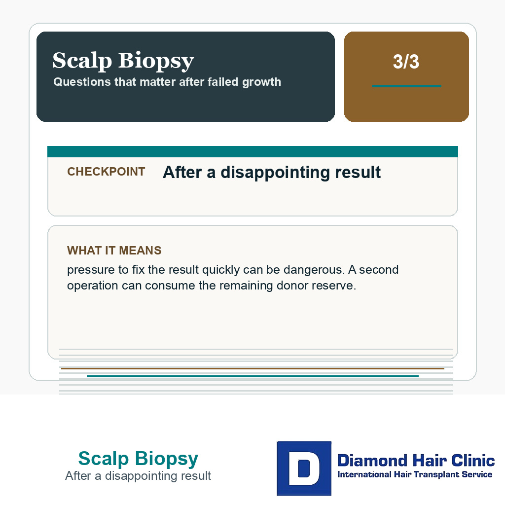
After a disappointing result, pressure to fix the result quickly can be dangerous. A second operation can consume the remaining donor reserve. In a poor hair transplant result and repair discussion, the responsible question is not “how many grafts can we add?” It is “why did this happen, and is the scalp safe to treat again?”
The 3 slides below split this section into one practical point per image. Swipe sideways, use the arrows to move one slide at a time, or use the numbered controls under the image to jump to a specific slide.


Checks that come before biopsy
Before biopsy is discussed, the history should be clear. I need to know when the hair loss started, whether it was sudden or gradual, and whether there is burning, itching, pain, scaling, bumps that look like acne, redness, or tenderness. Medication history also matters, including minoxidil, finasteride, dutasteride, isotretinoin, steroids, autoimmune medicines, or treatments used to quiet inflammation.
The scalp also needs close examination. Trichoscopy can show miniaturization, broken hairs, scale, perifollicular redness, loss of follicular openings, pustules, or signs that the donor area is not stable. Conditions such as folliculitis and hair transplant planning, seborrheic dermatitis before hair transplant, and scalp psoriasis and hair transplant surgery can all change timing even when biopsy is not needed.
Sometimes blood work, medication history, trichoscopy, or a dermatology diagnosis answers the question without biopsy. Biopsy becomes useful when the remaining uncertainty is at tissue level, such as whether there is scarring, active inflammation, follicle loss, or a mixed pattern that cannot be judged safely from the surface.
Biopsy location matters
A biopsy is only as useful as the question it is trying to answer and the area sampled. In many hair loss conditions, the most useful sample is not from completely smooth scarred skin or completely normal skin. The dermatologist often chooses an active edge where there are still follicles and where redness, scale, symptoms, or disease activity may be present.
A request to “just do a biopsy” is not enough. The sampling site, punch size, direction of sectioning, and pathology request can all affect the usefulness of the result. The pathologist also needs the clinical question, such as scarring versus loss that does not scar, active inflammation versus inactive change, or miniaturization versus mixed disease.

A scalp biopsy is more useful when the sampled area matches the diagnostic question, not when it is chosen randomly.
The limitation should be understood before the sample is taken. Biopsy can miss activity if the wrong area is sampled. It can show mixed findings. It can confirm a diagnosis but still not prove that surgery is safe today. The small biopsy site also leaves a wound that needs healing and should be noted during future planning so it is not confused with transplant scarring.
A useful biopsy result changes the decision. It does not simply approve surgery.
Biopsy results can change the plan
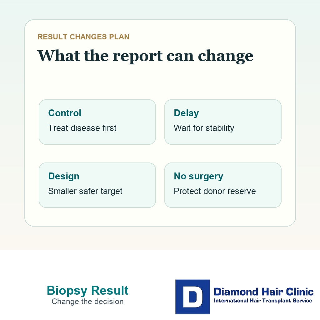
If biopsy supports ordinary androgenetic alopecia and the donor area is strong, the surgical conversation may become clearer. That does not make surgery automatic. It only removes one major uncertainty. The plan still has to return to donor capacity, hairline design, graft distribution, future progression, and expectations.
If biopsy shows scarring alopecia or active inflammation, the plan may change completely. Surgery may need to wait until dermatology treatment controls the disease and the scalp shows stability over time. The target may become smaller. A test area may be discussed. In some cases, surgery may be rejected because the risk of poor growth, disease recurrence, or further donor loss is too high.
If biopsy shows a mixed pattern, the answer may be more nuanced. A person can have androgenetic alopecia and inflammation at the same time. The surgical decision has to respect both problems. Transplanting only because one part of the diagnosis is surgical can ignore the part that makes surgery risky.
Questions to ask before booking surgery
If biopsy has been suggested, ask what diagnosis is being considered, why the current examination is not enough, who will take the sample, where it will be taken, what pathology review is needed, and how the result will change the surgical decision. If nobody can explain what decision depends on the biopsy, the test may not be well framed.
If biopsy has already been done, bring the full written pathology report, not only a verbal summary such as “it was normal.” I review whether the report mentions miniaturization, inflammation, scarring change, follicle loss, mixed findings, the biopsy site, sectioning method, or a limitation in the sample. Those details can change the surgical conversation.
If biopsy has not been suggested but you have burning, itching, redness, scaling, patchy loss, donor thinning, diffuse shedding, a previous failed transplant, or very different opinions from clinics, ask whether a dermatology diagnosis should come before surgery. That may or may not mean biopsy. It does mean the uncertainty should be named.
When advice conflicts, a second opinion before hair transplant surgery should not be just another graft quote. It should answer whether the diagnosis, donor area, scalp condition, and expectations support surgery. Do not let a clinic turn uncertainty into a transplant date. The useful order is diagnosis first, scalp stability second, surgical planning third.